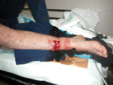
Trauma can lead to muscle ischaemia. Oedema occurs from release of free radicals increasing vascular permeability as blood flow is re-established. This results in muscle swelling.
In rigid compartments this can lead to compartment syndrome.
Muscle swelling leads to venous outflow obstruction, decreased arterial flow, further muscle ischaemia and ultimately infarction and necrosis.
The classic site is the calf, or the shin, due to its inexpansible fascial compartments; however, it can occur in the forearm.
Immediate intern managementAttend patient and assess. Suspect the diagnosis in any patient complaining of increasing pain in a limb post injury or surgery.
If in a well supported clinical environment, you may wish to discuss the situation before splitting a plaster or removing a bandage. |
A tonometer can be created if one is not available by connecting an arterial pressure transducer to a primed arterial line tube and a spinal needle. Once correctly zeroed, the spinal needle can be inserted into a tissue compartment to record the pressure in the compartment.
Measure rules of thumb:
If diagnosis is obvious (uncommon):
If diagnosis is unclear:
There are four compartments in the calf: peroneal, anterior, posterior superficial and posterior deep. The pressures in each compartment can be measured using a tonometer.
If doubt remains or diagnosis has been made ® urgent four-compartment fasciotomy.